Magnetic Resonance Imaging, or MRI, is one of the most advanced diagnostic tools available in modern medicine. Unlike X-rays or CT scans, which use ionizing radiation, MRI uses powerful magnets and radio waves to generate detailed images of the inside of the body. This non-invasive imaging technique is incredibly versatile and is used across a range of medical specialties to diagnose and monitor conditions affecting the brain, spine, muscles, joints, and other organs.
In this blog, we’ll explore how MRI works, when it’s commonly used, and what patients can expect during the procedure.
How MRI Works
MRI is based on the principles of magnetism and radio waves. Here’s a simplified breakdown of the process:
Magnetic Fields: The MRI machine generates a powerful magnetic field that temporarily realigns hydrogen atoms in the body. Since the human body is largely made up of water, and water is composed of hydrogen and oxygen atoms, the MRI machine focuses on the hydrogen atoms.
Radio Waves: Once the hydrogen atoms are aligned, the MRI machine sends a pulse of radio waves into the body. This pulse knocks the hydrogen atoms out of alignment, and when they return to their original state, they release energy.
Signal Detection: The MRI scanner detects the energy released by the hydrogen atoms and uses this data to create highly detailed images of the tissues in the body. The signals from different tissues (such as muscles, fat, or organs) produce varying results, allowing the machine to differentiate between types of tissues.
Computer Processing: The signals are sent to a computer, which processes the data into cross-sectional images. These images can be viewed in multiple planes (e.g., axial, sagittal, coronal) and even reconstructed into 3D models for better visualization.
When Is MRI Used?
MRI is used to diagnose and monitor a wide variety of health conditions. Here are some common indications for an MRI:
1. Neurological Conditions
MRI is often used to assess the brain and spinal cord due to its ability to provide high-resolution images of soft tissues. Some conditions where MRI is particularly useful include:
- Brain tumors: MRI can help detect tumors, monitor their size, and plan treatments.
- Multiple sclerosis (MS): MRI is essential for diagnosing MS, as it can identify characteristic lesions in the brain and spinal cord.
- Stroke: MRI can detect areas of the brain affected by a stroke and assess the extent of damage.
- Spinal cord injuries: MRI helps evaluate spinal cord trauma, herniated discs, and other spinal conditions.
2. Musculoskeletal and Joint Issues
MRI is commonly used in orthopedic medicine to evaluate joints, muscles, tendons, and ligaments. It is particularly useful in diagnosing soft tissue injuries, such as:
- Torn ligaments or tendons: MRI can detect tears in soft tissues, like the ACL in the knee or rotator cuff in the shoulder.
- Herniated discs: MRI is highly effective in identifying disc herniations in the spine that may cause nerve compression.
- Bone and joint abnormalities: MRI can help diagnose conditions like arthritis, joint degeneration, and infections affecting bones and joints.
3. Cardiovascular Imaging
MRI is increasingly being used to assess heart conditions, such as:
- Cardiomyopathy: MRI helps assess the condition of the heart muscle in patients with cardiomyopathy (heart muscle disease).
- Coronary artery disease: It can evaluate blood vessels, detecting blockages or other abnormalities that might lead to heart attacks.
- Congenital heart defects: MRI is valuable in identifying heart defects, particularly in children and adults born with these conditions.
4. Cancer Diagnosis and Staging
MRI is often used to evaluate the size, location, and extent of tumors, especially in soft tissues. For example:
- Breast cancer: MRI is sometimes used to supplement mammography in women with dense breasts or those at high risk for breast cancer.
- Prostate cancer: MRI helps guide biopsies and monitor the progression of prostate cancer.
- Liver, kidney, and other cancers: MRI can help detect and stage cancers affecting soft tissues or organs that are harder to see on other imaging tests.
5. Abdominal and Pelvic Imaging
MRI is helpful in assessing organs like the liver, kidneys, and reproductive organs. Common uses include:
- Liver diseases: MRI can assess liver conditions, including cirrhosis, fatty liver disease, or tumors.
- Pelvic conditions: MRI can evaluate conditions affecting the uterus, ovaries, and prostate, such as fibroids, endometriosis, or cancer.

What to Expect During an MRI
If your doctor has recommended an MRI, it’s important to understand what to expect before, during, and after the procedure.
Before the MRI
- Preparation: In most cases, no special preparation is required. However, you may be asked to avoid food or drink for a few hours if the MRI is being performed on your abdomen or pelvis.
- Clothing: You will likely be asked to change into a hospital gown and remove any metal objects, such as jewelry, watches, and hearing aids. Metal can interfere with the magnetic field and affect image quality.
- Screening for Implants: Because of the strong magnetic field, MRI is not suitable for patients with certain metal implants (e.g., pacemakers, cochlear implants, metal stents). You will be asked about any previous surgeries or implants before the procedure.
During the MRI
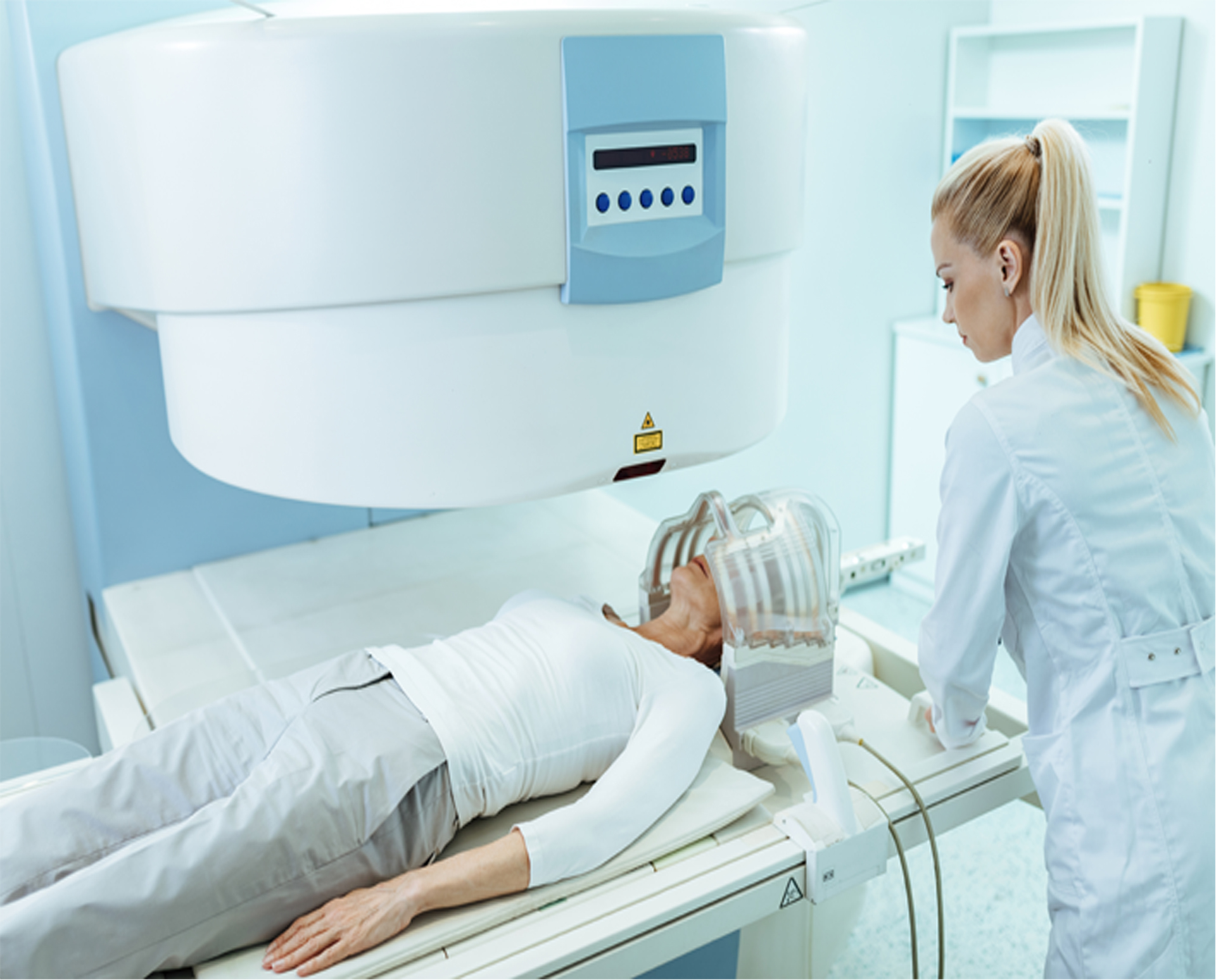
- Positioning: You will be asked to lie down on a moveable table. For certain exams (such as brain or spinal MRI), you may need to be positioned carefully to ensure accurate images.
- The MRI machine: The table will slide into the MRI machine, which is a large, cylindrical magnet. Some machines are “closed,” meaning you’ll be enclosed inside the tube, while others are “open” or have a wider opening, which may be less claustrophobic for some patients.
- Noise: MRI machines can be quite noisy, producing loud banging or tapping sounds. You may be given earplugs or headphones to reduce the noise.
- Duration: The MRI scan itself typically takes 20 to 60 minutes, depending on the type of scan. You’ll be asked to remain as still as possible to ensure clear images.
- Contrast Agents: In some cases, a contrast dye (gadolinium) may be injected into your veins to enhance the images. This is especially common for scans of the brain, spinal cord, and blood vessels.
After the MRI
- Results: Once the MRI is complete, the images will be reviewed by a radiologist, who will send a report to your doctor. Your doctor will then discuss the results with you.
- Recovery: In most cases, there’s no recovery time needed after an MRI. You can resume your normal activities immediately unless you received sedation or contrast dye, in which case your doctor may give you additional instructions.
Benefits and Limitations of MRI
Benefits:
- Non-invasive and radiation-free: MRI does not involve ionizing radiation, making it safer for repeated use, especially in sensitive populations like children and pregnant women.
- Excellent soft tissue imaging: MRI provides high-resolution images of soft tissues, which makes it particularly effective for imaging the brain, muscles, joints, and organs.
- Detailed and accurate: MRI can detect subtle abnormalities that might be missed by other imaging techniques.
Limitations:
- Cost and availability: MRI machines are expensive, and not all healthcare facilities have access to them. The procedure can also be more costly than other imaging techniques like X-rays or ultrasound.
- Claustrophobia: Some patients may experience anxiety or discomfort from being in the enclosed MRI machine, though open MRI machines are available at some facilities.
- Time-consuming: MRI scans can take longer than other imaging methods, which may be challenging for patients who have difficulty staying still or need to complete multiple scans.